Primary extraosseous Ewing sarcoma of the lung in children
Nidal Alsit 1, Clara Fernandez 2, Jean Luc Michel 3, Linda Sakhri 4, Audrey Derouet 5 and Augustin Pirvu 6
1 Department of Thoracic, Vascular, and Cardiac Surgery, University Hospital Felix Guyon, Reunion, France
2 Department of Pathological Anatomy, University Hospital Felix Guyon, Reunion, France
3 Department of Pediatrics Surgery, University Hospital Felix Guyon, Reunion, France
4 Department of Onco-pneumology, University Hospital Grenoble, France
5 Department of Pediatrics, University Hospital Felix Guyon, Reunion, France
6 Department of Thoracic, Vascular, and Endocrine Surgery, University Hospital Grenoble, France
Correspondence to: Augustin Pirvu. Email: pb_augustin@yahoo.com; apirvu@chu-grenoble.fr
The report in this article of the treatment the patient received is incorrect. Three of the authors (Jean Luc Michel, Clara Fernandez and Audrey Derouet) were unaware that their names had been added to the author list.
The three remaining authors (Nidal Alsit, Linda Sakhri and Augustin Pirvu) were not involved with the treatment of the patient.
Abstract
We report a case of primary extraosseous Ewing sarcoma (EES) of the lung in a four-year-old child. In the literature, there are only a few case reports of EES located in the thorax.
A Retraction for this article has been published in 2013 ecancer 7 332.
Keywords: lung tumor in children, extraosseous Ewing sarcoma
Background
Extraosseous Ewing sarcoma (EES) is an uncommon malignant neoplasm in which pulmonary localisation is exceptionally rare.
Case Report
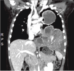
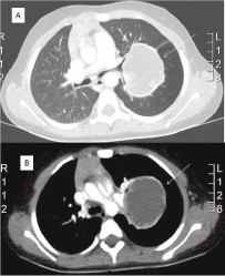
A four-year-old girl, without any medical history, was referred to our department for a lung mass (Figure 1). An initial thoracic computed tomography (CT) scan revealed a large cystic tumour in the middle of the left lung (Figure 2). The diagnosis of intrathoracic EES was made by punction under CT control. The patient was subsequently treated with six chemotherapy courses (vincristin, ifosfamid, doxorubicin, and etoposide).

Figure 1: CT scan: coronal reconstruction of a left lung mass (arrow)
At the end of chemotherapy, after a negative search for metastasis, we performed a radical resection, which consisted of a left pneumonectomy. The pathologic examination confirmed the need for a complete resection and also confirmed the initial diagnosis. During an interdisciplinary meeting, it was decided that postoperatively the patient would receive seven courses of chemotherapy (vincristin, actinomycin, and ifosfamid) without radiotherapy. The patient is currently receiving postoperative chemotherapy.
Discussion
In our case, the patient was a four-year-old girl, which was unusually young when compared with the previously reported cases [1–6]. The most common CT finding of EES reported is a heterogeneous mass [1–3], but in the present case, the CT showed the EES as a cystic structure.
The chemotherapy treatment was done according to the Eurowing 99 protocol and was also followed by a very aggressive surgery justified by the size, location, as well as the aggressive character of the tumour [2–5]. According to most of the authors, this kind of tumour should be resected as an attempt to obtain complete control of the disease [1–6].

Figure 2: CT scan: (A) cystic tumour in the middle of left lung between the superior and the lower bronchus with maximum diameter of 10/8 cm (arrow); (B) close contact of the tumour (arrow) with the left pulmonary artery
Conclusion
Intrathoracic EESs are extremely rare and complex conditions requiring a pluridisciplinary collaboration. This case highlights the importance of preoperative evaluation and strategy in aggressive tumours.
Conflicts of interest
The authors declare that there are no conflicts of interest that could be perceived as prejudicing the impartiality of the research reported.
References
1. Siddiqui MA, Akhtar J, Shameem M, Baneen U, Zaheer S and Shahid M (2011) Giant extraosseous Ewing sarcoma of the lung in a young adolescent female–a case report Acta Orthop Belg 77(2) 270–3 PMID: 21667743
2. Halefoglu AM (2012) Extraskeletal Ewing’s Sarcoma Presenting as a Posterior Mediastinal Mass Arch Bronconeumol [epub ahead of print] DOI: 10.1016/j.arbres.2012.02.020 PMID: 22575810
3. Xie CF, Liu MZ and Xi M (2010) Extraskeletal Ewing’s sarcoma: a report of 18 cases and literature review Chin J Cancer 29(4) 420–4 DOI: 10.5732/cjc.009.10402 PMID: 20346219
4. Hancorn K, Sharma A and Shackcloth M (2010) Primary extraskeletal Ewing’s sarcoma of the lung Interact Cardiovasc Thorac Surg 10(5) 803–4 DOI: 10.1510/icvts.2009.216952 PMID: 20067987
5. Lee YY, Kim do H, Lee JH, Choi JS, In KH, Oh YW, Cho KH and Roh YK (2007) Primary pulmonary Ewing’s sarcoma/primitive neuroectodermal tumor in a 67-year-old man J Korean Med Sci 22 S159–63 DOI: 10.3346/jkms.2007.22.S.S159 PMID: 17923745
6. Kara IO, Gonlusen G, Sahin B, Ergin M and Erdogan S (2005) A general aspect on soft-tissue sarcoma and c-kit expression in primitive neuroectodermal tumor and Ewing’s sarcoma. Is there any role in disease process? Saudi Med J 26(8)1190–6 PMID: 16127511


