Bulky palpebral Merkel cell carcinoma of the eyelid
Dario Alvaro Rueda1,a, Cecilia Schweitzer2, Di Nisio Lorena2, María Laura Piccoletti3, Nicolas Torressi1, Víctor Acevedo1 and Silvia Ferrandini1
1Clinical Oncology Division, Hospital de Clínicas José de San Martin (UBA), Buenos Aires 1120, Argentina
2Oculoplastic Surgery Department, Hospital de Clínicas José de San Martin (UBA), Buenos Aires 1120, Argentina
3Otorhinolaryngology Department, Hospital de Clínicas José de San Martin (UBA), Buenos Aires 1120, Argentina
ahttps://orcid.org/0009-0005-5657-8463
Abstract
Merkel cell carcinoma (MCC) is a rare and aggressive tumour of the skin, characterised by a high rate of local recurrence and lymph node involvement. We present the case of a 58-year-old woman who developed a 5-cm tumour on the right lower eyelid, leading to ocular occlusion. Magnetic resonance imaging revealed an exophytic lesion in the right orbit, and a biopsy confirmed the diagnosis of MCC. After complete surgical resection and cervical emptying, the patient was treated with adjuvant radiotherapy. The final diagnosis was MCC, stage pT3 pN1 M0. The periocular location and tumour size were determinants in the treatment decision.
Keywords: Carcinoma, Merkel cell/therapy, skin neoplasms/etiology, male, neoplasm staging
Correspondence to: Dario Alvaro Rueda
Email: darioalvarorueda@gmail.com
Published: 17/07/2025
Received: 28/10/2024
Publication costs for this article were supported by ecancer (UK Charity number 1176307).
Copyright: © the authors; licensee ecancermedicalscience. This is an Open Access article distributed under the terms of the Creative Commons Attribution License (http://creativecommons.org/licenses/by/4.0), which permits unrestricted use, distribution, and reproduction in any medium, provided the original work is properly cited.
Introduction
Merkel cell carcinoma (MCC) is a rare and aggressive tumour of the skin, characterised by a high rate of local recurrence and nodal involvement. Risk factors include advanced age, male gender, white race and immunocompromised status (such as HIV infection, organ transplantation or B-cell neoplasms) [1–3].
MCC was first described in 1972 by Cyril Toker referring to it as ‘trabecular carcinoma of the skin’ [4].
The most frequent location is in the head and neck area (43%) [1–3]. Approximately 65% of cases are diagnosed in localised phase, 26% present locoregional involvement and 8% are metastatic [2].
Locoregional recurrence rates range from 30% to 60%, and up to one-third of patients develop distant metastases [5].
Clinical case
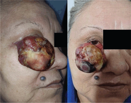
A 58-year-old woman with no relevant medical history presented with a progressively enlarging tumour on the right lower eyelid first noticed 3 months prior. The lesion reached approximately 5 cm in diameter, leading to complete occlusion of the right eye and generating significant anxiety for the patient, primarily due to aesthetic concerns. Despite its size, the lesion was painless and did not compromise vision (Figure 1).
Clinically, the tumour appeared as an exophytic, firm-elastic, lobulated mass with a reddish-violet coloration and a yellowish shiny surface, superficial vascularisation, and a necrotic area in the lower portion. The borders were well-defined, with no ulceration or visible discharge.
A biopsy revealed proliferation of neoplastic cells showing diffuse and intense positivity for CK AE1/AE3, cytokeratin 20, and chromogranin, and negativity for CD20 and CD3. The Ki-67 proliferation index was 90%. These findings confirmed the diagnosis of MCC.
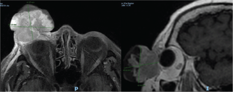
Magnetic resonance imaging (MRI) with gadolinium demonstrated a large exophytic lesion in the right orbit, located in the preseptal region and arising from the lower eyelid. After intravenous contrast administration, the lesion showed heterogeneous enhancement and was associated with ipsilateral enophthalmos, consistent with mass effect. The lesion measured approximately 45 × 38.7 mm in the axial plane (Figure 2).
A contrast-enhanced computed tomography (CT) scan of the neck, thorax, abdomen, and pelvis revealed no distant metastases, although infracentimetric right cervical lymph nodes were observed.
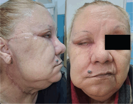
Complete resection of the tumour was performed, along with a right cervical lymph node dissection and parathyroidectomy (Figure 3). Intraoperatively, the tumour appeared well delimited, with no evidence of bony or intraorbital invasion. A wide excision with 1 cm clinical margins was achieved. Following tumour resection, a total right parotidectomy was performed with preservation of the facial nerve, confirming intraparotid metastatic involvement with macroscopically free margins. A selective neck dissection of levels I, II, and III was also carried out on the ipsilateral side. The skin and soft tissue defect of the cheek was reconstructed using a Mustardé flap, achieving adequate eyelid closure.

Figure 1. Frontal and oblique clinical photographs showing a large exophytic tumour originating from the right lower eyelid. According to the TNM classification for MCC, this corresponds to a cT2 lesion (greater than 2 cm but less than 5 cm). Based on the TNM system for eyelid carcinoma, it qualifies as cT3c (tumour >3 cm involving the full thickness of the eyelid).

Figure 2. Contrast-enhanced MRI of the brain and orbits. A large exophytic mass is seen in the right orbit, located in the preseptal region and arising from the lower eyelid. The lesion shows heterogeneous contrast enhancement and is associated with ipsilateral enophthalmos due to mass effect. Its axial dimensions are approximately 45 × 38.7 mm.

Figure 3. Frontal and lateral postoperative clinical photographs. The images show the surgical scar following resection of the MCC and the right cervical lymphadenectomy.
The histopathological analysis described a 6 × 6 × 4 cm tumour of the right lower eyelid with areas of necrosis and hemorrhage. There was evidence of vascular emboli and perineural invasion. Surgical margins were free of tumour. The right cervical dissection showed no metastases in the 27 lymph nodes analysed. However, 2 of 3 nodes in the area adjacent to the parotid gland demonstrated carcinomatous infiltration with vasculolymphatic emboli, but without capsular rupture. The final pathological staging was pT3 pN1, consistent with MCC.
Given the tumour location in the head and neck region and the large size, adjuvant intensity-modulated radiotherapy was initiated in the surgical bed and regional lymphatic drainage areas.
The patient was followed clinically and radiologically for 15 months, with no evidence of local or distant recurrence at the last follow-up. Eyelid function was preserved, with resolution of postoperative residual facial nerve paresis, and satisfactory cosmetic adaptation of the reconstructive graft.
Discussion
Although the face is a frequent location of MCCs, eyelid tumours account for only 2.5% of cases [3]. Interestingly, women are about twice as likely as men to develop an MCC on the eyelids [3, 4].
Within eyelid tumours, the upper eyelid is the most commonly affected, as opposed to basal cell and squamous cell carcinomas [3].
The extent of disease at diagnosis, lymph node involvement (the main predictor of survival), lymphovascular invasion and infiltrative growth pattern are poor prognostic factors, as is the expression of p63 [2].
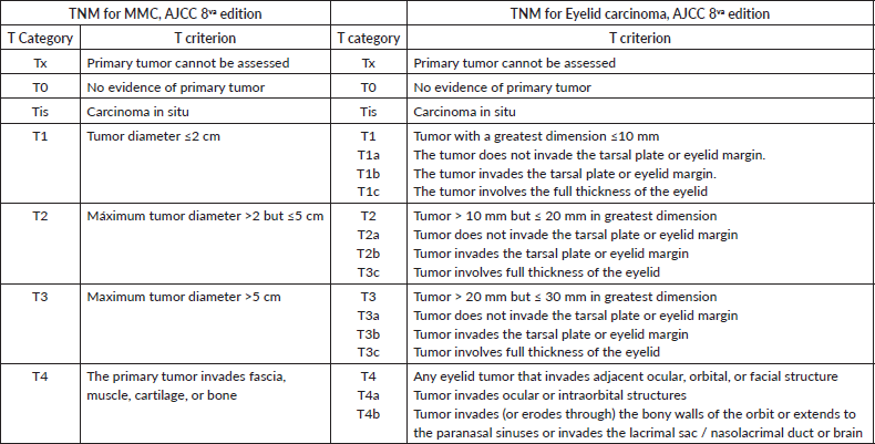
Tumour size at presentation is also a crucial factor in MCC of the periocular region and eyelids. The eighth edition (2017) of the American Joint Committee on Cancer (AJCC) Tumor, Node, Metastasis (TNM) system uses a tumour size of 2 cm or less to classify all lesions as T1, although no specific guidelines are provided for eyelid lesions. Specific staging of eyelid carcinoma correlates better with disease-free survival in these cases, subdividing lesions into T1 for tumours smaller than 5 mm, T2a for those between 5 and 10 mm, and T2b for tumours 10 to 20 mm (Table 1) [1, 4–6].
As for treatment, in patients with localised disease, surgical resection with negative margins of 1–2 cm radiated to the tumour is the treatment of choice [1, 2]. In tumours of the periocular region, wide local excision with 5 mm margins is recommended [3, 4, 7].
Table 1. Comparison between T category of MCC TNM and eyelid carcinoma TNM1,6

For patients at high risk of local recurrence after resection of the primary tumour, adjuvant radiotherapy is suggested. High-risk features include primary tumours of ≥1 cm, positive or close surgical margins, lymphovascular invasion, head and neck location, and immunocompromised status [1, 2].
Although there are no randomised trials supporting the role of adjuvant radiotherapy, observational studies suggest that it decreases the rate of locoregional recurrence and improves overall survival [1]. It is recommended to initiate radiotherapy within 8 weeks after surgery [1, 2].
There is no evidence for the use of chemotherapy or immunotherapy with checkpoint inhibitors in the adjuvant setting. Immune checkpoint inhibitors (ICIs) such as avelumab are indicated in metastatic or unresectable disease [1, 2].
Although neoadjuvant is not a standard of care, a phase I/II study of 39 patients with stage IIA-IVA MCC demonstrated pathologic complete responses close to 50% with two doses of Nivolumab 240 mg. This option may be an alternative mainly in patients whose surgery is borderline, always in selected cases and evaluated in a committee of experts [2, 8].
Conclusion
MCC of the eyelid is a rare but aggressive entity, with prognosis influenced by anatomic location and tumour size. In this case, surgical excision followed by adjuvant radiotherapy was the treatment of choice due to the head and neck location, the significant size of the lesion, and the risk of locoregional recurrence. Timely and multidisciplinary management is key to improve the prognosis in this type of patient.
Conflicts of interest
The authors declare that there is no conflicts of interest in the preparation of this manuscript.
Funding
No funding was received for this study.
Author contributions
All authors have contributed to the preparation of the manuscript.
References
1. Schmults C, Blitzblau R, and Aasi SZ, et al (2023) Merkel cell carcinoma Clin Pract Guidelines Oncol [Internet] [https://www.nccn.org/home/member] Date accessed: 14/09/24
2. Anabella L, Pedro B, and Florencia C, et al (2024) Skin tumors Current Recommendations for Oncologic Treatment of the Argentine Society of Clinical Oncology (Buenos Aires: AAOC)
3. North VS, Habib LA, and Yoon MK (2019) Merkel cell carcinoma of the eyelid: a review Surv Ophthalmol 64(5) 659–667 https://doi.org/10.1016/j.survophthal.2019.03.002 PMID: 30871952
4. Merritt H, Sniegowski MC, and Esmaeli B (2014) Merkel cell carcinoma of the eyelid and periocular region Cancers (Basel) [Internet] 6(2) 1128 Date accessed: 07/07/24 https://doi.org/10.3390/cancers6021128 PMID: 24821131 PMCID: 4074820
5. Sniegowski MC, Warneke CL, and Morrison WH, et al (2014) Correlation of American joint committee on cancer T category for eyelid carcinoma with outcomes in patients with periocular merkel cell carcinoma Ophthalmic Plast Reconstr Surg 30(6) 480–485 https://doi.org/10.1097/IOP.0000000000000153 PMID: 24841735
6. Ding S, Sagiv O, and Guo Y, et al (2019) Change in eyelid carcinoma T category with use of the 8th versus 7th edition of the American Joint Committee on Cancer: cancer staging manual Ophthalmic Plast Reconstr Surg [Internet] 35(1) 38–41 Date accessed: 17/09/24 https://doi.org/10.1097/IOP.0000000000001133
7. Herbert HM, Sun MT, and Selva D, et al (2014) Merkel cell carcinoma of the eyelid management and prognosis JAMA Ophthalmol 132(2) 197–204 https://doi.org/10.1001/jamaophthalmol.2013.6077
8. Topalian SL, Bhatia S, and Amin A, et al (2020) Neoadjuvant nivolumab for patients with resectable Merkel cell carcinoma in the CheckMate 358 trial J Clin Oncol [Internet] 38 2476–2787 https://doi.org/10.1200/JCO.20.00201 PMID: 32324435 PMCID: 7392746


